Skip to content
The human heart is a remarkable organ that beats approximately 100,000 times daily, pumping blood throughout the body to deliver oxygen and nutrients to our cells. However, like any other body part, the heart is vulnerable to disease and damage.
That’s where electrocardiograms (ECGs) come in. ECGs are a valuable diagnostic tool that can provide critical information about the heart’s health. In this article, we’ll delve into the world of ECGs and explore what they are, how they work, and what they can tell us about heart health. We’ll also discuss the procedure for performing an ECG, how to interpret ECG results, and what to do if ECG results are abnormal.
Whether you’re a healthcare professional or interested in heart health, this article will provide a comprehensive overview of ECGs and their importance in diagnosing and monitoring heart conditions.
What Is An Electrocardiogram (ECG)?
At its core, an electrocardiogram (ECG) is a non-invasive test that measures the heart’s electrical activity. This electrical activity is essential for the heart to beat and pump blood effectively throughout the body. By recording the electrical signals the heart produces, ECGs can provide valuable information about the heart’s structure, function, and rhythm.
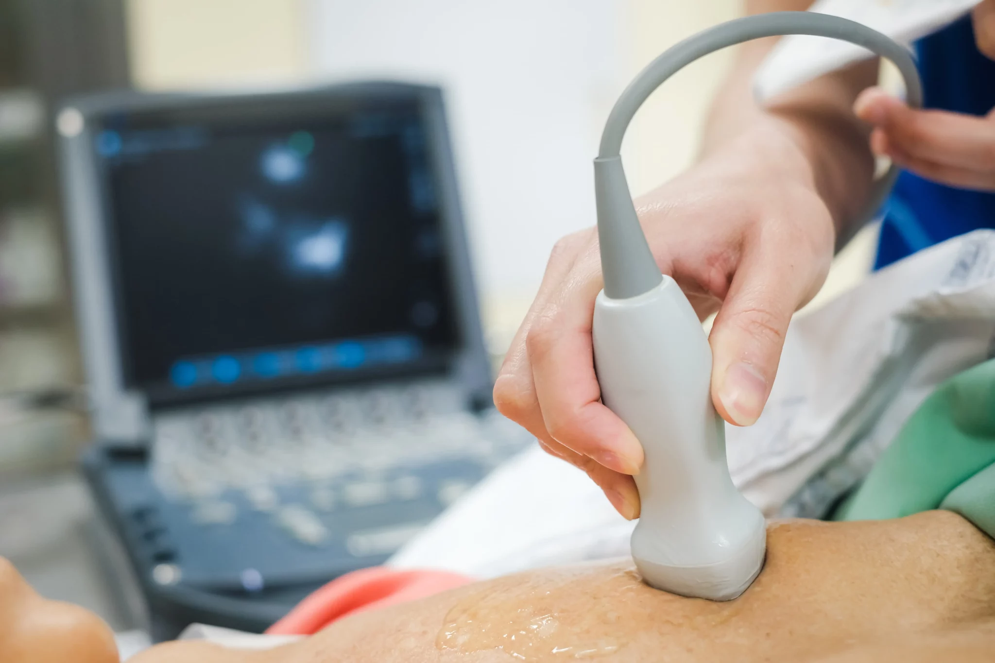
An ECG is performed using a machine that detects and amplifies the electrical signals produced by the heart. Electrodes are placed on the skin of the chest, arms, and legs, and these electrodes are connected to the ECG machine. The machine then records the heart’s electrical activity, which is displayed as a graph on paper or on a computer screen.
ECGs can be performed in a doctor’s office, clinic, or hospital, and they typically take between 5 and 10 minutes to complete. The test is painless, and it has no risks or side effects.
There are several different types of ECGs, including a resting ECG, which is performed while the patient is at rest, and a stress ECG, which is performed while the patient is exercising. Each type of ECG provides different information about the heart and can be useful in diagnosing other heart conditions.
What Can ECGs Tell Us About Heart Health?
Electrocardiograms (ECGs) are a powerful diagnostic tool that can provide critical information about heart health. ECGs measure the heart’s electrical activity and can detect abnormal patterns that may indicate underlying heart conditions.
Normal ECG patterns include a series of distinct waves and intervals representing the electrical signals generated by different heart parts as it contracts and relaxes. On the other hand, abnormal ECG patterns may indicate various heart conditions, ranging from mild to life-threatening.
- Detect arrhythmias, which refer to an irregular heartbeat caused by various factors such as heart disease, high blood pressure, and certain medications. ECGs can detect arrhythmias by measuring the timing and strength of the electrical signals generated by the heart.
- Detect signs of heart attack, which occurs when blood flow to the heart is blocked, causing damage to the heart muscle. During a heart attack, the heart’s electrical activity can change rapidly, and ECGs can detect these changes, helping healthcare professionals diagnose and treat the condition.
- Detect signs of heart failure occur when the heart cannot pump enough blood to meet the body’s needs. ECGs can detect signs of heart failure by measuring the heart’s electrical activity and identifying any abnormalities.
- Identify valve problems and congenital heart defects. ECGs can provide critical information about the heart’s structure, function, and rhythm, helping healthcare professionals diagnose and monitor heart conditions.
How Are ECGs Performed?
During an ECG, the patient lies down on a bed or table, and several small electrodes are attached to the skin on the chest, arms, and legs. These electrodes are connected to a machine that records the heart’s electrical activity and produces a graph called an electrocardiogram.
Patients are asked to remain still and avoid talking or moving during the procedure to get the most accurate reading. Healthcare professionals may also ask patients to take deep breaths or hold their breath briefly to help stimulate changes in the heart’s electrical activity.
ECGs are painless and generally considered safe. However, patients with certain medical conditions, such as pacemakers or implanted defibrillators, may need to take special precautions or avoid the procedure altogether. Patients should always inform their healthcare provider of any medical conditions or medications they are taking before undergoing an ECG.
After the procedure, healthcare professionals analyze the electrocardiogram to identify abnormalities or irregularities in the heart’s electrical activity. Further testing, such as a stress test or echocardiogram, may be recommended if a patient’s ECG indicates a potential heart condition.
Interpreting ECG Results
Interpreting electrocardiogram (ECG) result is a complex process that requires specialized training and expertise. The ECG graph is made up of several components, each of which provides valuable information about the heart’s electrical activity.
The first component is the P wave, which represents the electrical activity of the atria or the heart’s upper chambers. A normal P wave indicates the electrical signal travels smoothly through the atria.
The next component is the QRS complex, which represents the electrical activity of the ventricles or the heart’s lower chambers. A normal QRS complex indicates that the electrical signal travels smoothly through the ventricles.
The final component is the T wave, which represents the repolarization, or recovery, of the ventricles. A normal T wave indicates that the ventricles recover appropriately after each heartbeat.
ECG results should always be interpreted in the context of the patient’s overall health and medical history. A single abnormal ECG does not necessarily indicate a serious heart condition; further testing may be needed to confirm a diagnosis.
Patients should always discuss their ECG results with their healthcare provider to fully understand the implications for their heart health. With the help of ECGs and other diagnostic tools, healthcare professionals can diagnose and treat heart conditions early, improving patient outcomes and quality of life.
Takeaway
If you are concerned about your heart health or have been advised by your healthcare provider to undergo an electrocardiogram (ECG), Roshal Health is here to help. Our state-of-the-art imaging facilities, highly trained technicians, and healthcare professionals can provide the care and expertise you need to monitor and manage your heart health.
Take your time – schedule your ECG appointment with Roshal Health today. Our team will work with you to ensure a comfortable and stress-free experience and provide the information and support you need to take charge of your heart health. Contact us now to learn more about our services and schedule your appointment.